Phase 1: my stem cells
For the past 6 months, I’ve been training myself to make stem cells. Of course, what does making stem cells entail? Some may have learned early on that your body stops producing stem cells except in your hippocampus and olfactory bulbs. My friends and family would ask why are you doing this? Or more importantly, when will you stop going to school? Well, I hope this post will answer all of your inquiries.
First I want to provide some background into stem cells and give attribution to the amazing feat that deduced a complex process of literally Benjamin Button-ing adult cells back to their nascent stem cell state. Around the mid-2000s, scientists figured out a way to take adult cells such as fibroblasts and later peripheral blood mononuclear cells (PBMC) and reprogrammed them into induced pluripotent stem cells (IPSC). After many passes through different combinations of transcription factors and oncogene, they deduced the necessary genes to reprogram human cells down to these main activators: KOS (polycistronic Klf4-Oct3/4-Sox2), Myc, Klf4 (because one is not enough) (see this amazing review of the history of stem cell historical discoveries Omole and Fakoya, 2018).
From what I hear, it’s one of the hardest cell culture protocols. So why am I going down this path of creating my own stem cells? Well because my masochistic tendencies love to explore the hardest but shortest path of modeling my own disease.
“Why couldn’t you just hire someone to do this for you?”
Well, science and labs are in short supply and even if I paid someone to make my stem cells — which you can — I am still short of modeling my rare inherited retinal disease. Stem cells are only a precursor or the first step to making a disease model after a patient’s specific gene mutation. The next step is to take stem cells and study the cells that best represent the disease. In my case, a retinal cell is the best way to study my retinal disease.
“Why do you need to study retinal cells from humans? Couldn’t you study this in animals?”
I wish I had the luxury to model my disease on animals. Here’s the catch, EYS does not exist in rodents. I repeat EYS gene is nowhere to be found in the rodent gene and this includes mice, rats, squirrels, and tree shrews (we’ve looked). But we have studied them in our favorite fish and invertebrate animal model, zebrafish and drosophila. Although we can study it, the EYS gene between humans and zebrafish shares about 33% similarity. This makes it hard to map the mutations that cause my disease from humans to fish and vice versa. Second, if I were to find a treatment using gene therapy and I had used a fish to model my disease, then I would have to design the therapy for the fish first and then I would have to go back and re-engineer that therapy to my gene. So given my conundrum, I needed to find a lab in the Bay Area that would study retinal diseases using patient-derived cells. In fact, I found professor Deepak Lamba (lab link) who models retinal diseases by making mini-retina clusters in a petri dish called retinal organoids. He works in reconstructing the retina morphologies from patient-derived stem cells so we have a better understanding of the mechanism behind the disease. If you wanted to know how I ended up in Deepak’s lab, check out this post here.
So I started my first cell culture in a little over a decade by grabbing a frozen aliquot of PBMCs (peripheral blood mononuclear cells) that have been collected from my blood draw back in June (boy, do I look like a freshman grad student, innocent and naive).

Back in June, I pose with my my blood draw and getting ready to get it processed in the lab.
I’ve attempted 2 times to reprogram the PBMC to IPSC and I’ve always stopped short 35 days after reprogramming to find either no colonies or small colonies that went nowhere (see the image). I’m telling you it’s very sad. They just refused to grow. What gives?
Day 34 and colonies were no more 16 cells.
Third time’s a charm, right? Well, the charm came from reprogramming fresh blood right after finishing up running some tests for ProEYS Natural History Clinical Trial that I am in (see link to learn more). And yes, it made a HUGE difference. Because in less than 13 days, I got colonies so big that I started picking them early. So far every 4-6 days, I kept picking colonies and started my passages. Today I am on passage 3 of taking undifferentiated IPSC colonies and culturing them so that we only have undifferentiated IPSC colonies. This guarantees that you have cells that are most likely stem cells and not cells that have been differentiated into something.
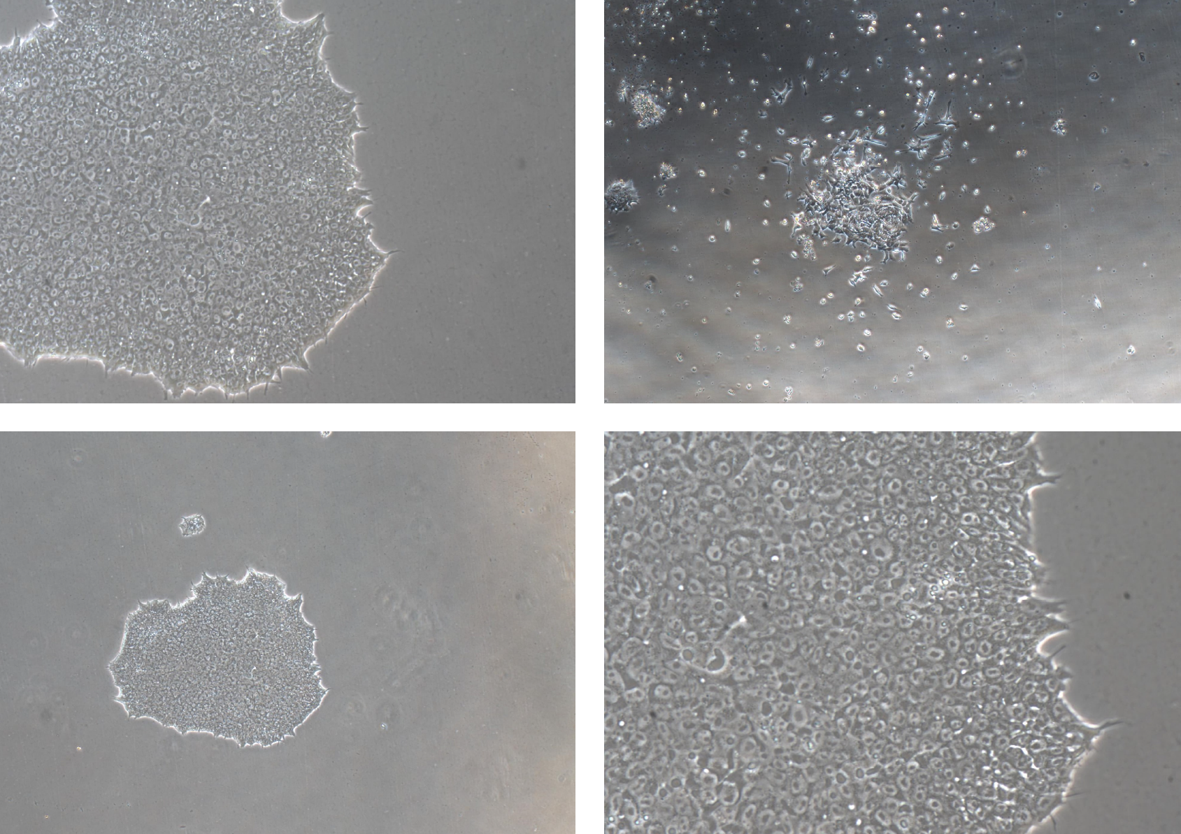
Stem cells packed into a single colony compared to very spaced out differentiated (non-stem cells). Top left: 10x magnification of colonies. Bottom left: 4x magnification to see entire colony. Bottom right: 20x magnification to see packed cells. Top right: non-stem cells not packed densely into a colony.
Now that I am finishing up the first phase, the second phase of making mini-retinas (retinal organoids) will start as soon as the first of February. This second phase is long and arduous and requires the forethought of making enough organoids that can survive more than 150 days, which is when photoreceptors start to appear. It is then that we hypothesize that EYS protein will express near the base of the outer segment as seen below in macaque retinas.

EYS shown in green is expressed at the base of the outer segment of both rod and cone photoreceptors. You can see the higher expression of EYS in the outer segment of blue cone receptors, which is co-stained with blue opsin (Yu et al., 2016).
Then we beg to ask, what does EYS do that is so important in the retina? What do the mutations do to my retina that renders me to lose my vision in my 30s but somewhat perfectly intact until my adult life? Will I even see any differences in the mini-retinas between my mutations vs. healthy ones? I hope to answer some of these questions in Phase 2.
If I could get any help in this journey, it is to connect to experts and collaborators who are interested in the following unknowns:
The preclinical requirements of getting treatments prepared for clinical trials. How does an N-of-1 (1-to-N) toxicology screen look like for someone who is screening treatments on retinal organoids? Is organoid or cell survival enough?
Are there any therapies worth testing that can preserve and protect existing photoreceptors? It could be anything from NAC (n-acetyl-cysteine) to Antabuse and metformin.
What are some worthy experiments/questions besides protein/transcript expression in these organoids? Harder questions like how to search for other protein-protein binding interactions? How to target for each of the four different isoforms?
Until then, spread the word, the awareness of (1) rare diseases and how most of them are left unstudied, (2) those who are fortunate like me can find funding to research my own rare disease, and (3) there are pathways being paved now as I continue to help patients through Perlara. Never give up hope.